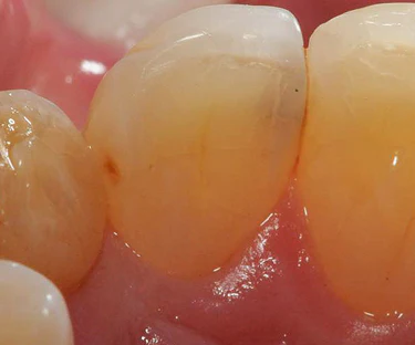
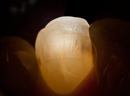
Cracked tooth diagnosis
Experience the Microlux 2: Your Essential Tool for Precise Diagnosis of Cavities and Fractures
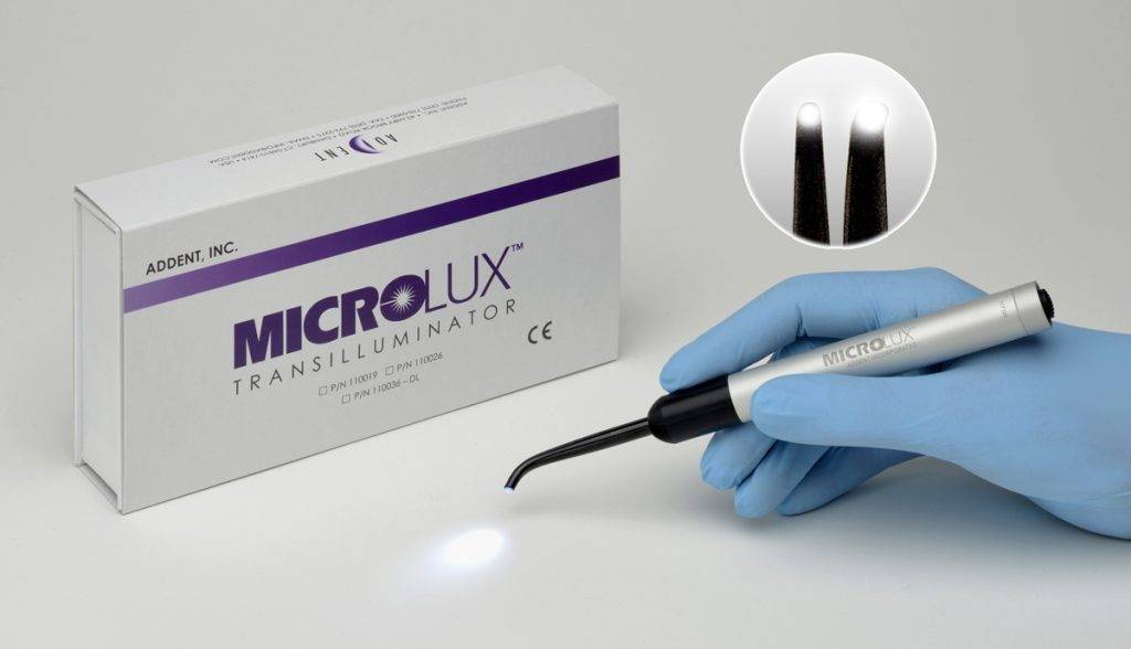
“Upgrade Your Practice with Microlux 2: The Compact Solution for Confident Diagnosis and Treatment Planning
Are you often uncertain about cracked teeth, torn between simple fixes and complex referrals? The Microlux 2 Transilluminator by AdDent offers a clear view, easing your decision-making process.
Crafted with dentist input, the Microlux 2 is a pocket-sized powerhouse, revealing intricate tooth damage at an affordable price. Its intuitive design requires just one finger for operation, featuring push-button controls and an anti-roll feature.
Equipped with various tips, this tool adapts to diverse dental needs:
- 2mm or 3mm lights aid in diagnosing anterior and posterior caries and fractures, with the 3mm tip also pinpointing root canals.
- The Proximal Caries tip targets posterior caries, while the Endo Lite tip locates root canals in the sulcus.
- The Perio-Probe attachment measures gingival recession at different depths.
- Compatible with all existing Mircolux attachments, its versatility enhances your practice.
Renowned dentist Dr. James R. Dunn hails the Microlux as indispensable for evaluating dental tissues, utilizing it in various procedures.
Many practitioners opt for multiple units, with its AA battery power and voltage regulator ensuring uninterrupted use. Dental Product Shopper Evaluators rated it 4.1 out of 5, citing its reliability and performance.
Elevate your practice with Microlux 2. Learn more here.
Clinical Technique
Example of Use:
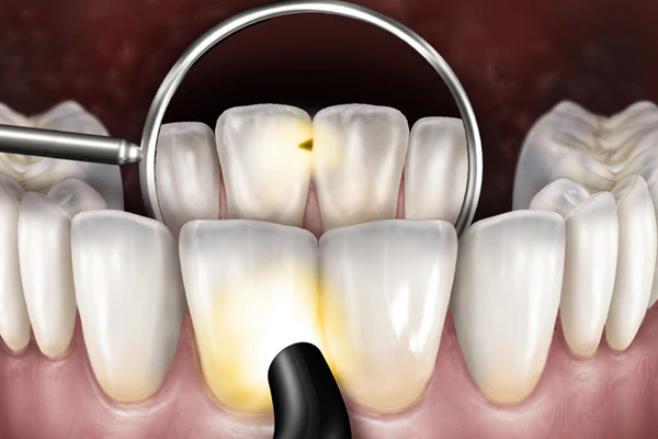
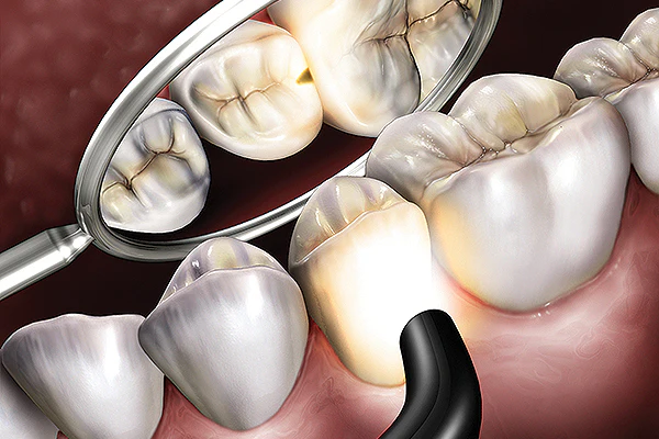
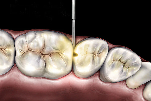
Anterior Caries: anterior caries can be visualized by placing the probe on the labio-cervical region of the tooth and examining from the lingual.
Posterior Caries: to visualize posterior proximal decay, place the probe on the cervical area of the tooth. Caries appear as a dark shadow on the occlusal surface.
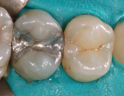
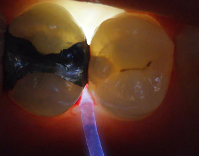
Posterior Caries: caries can also be detected using the Proximal Cariers fibre tip. Place the lighted fibre tip interproximally under the marginal ridge. View from the occlusal angle. This method often shows caries with high definition.
Identifying Proximal Posterior Caries
Photos courtesy Howard E. Strassler, DMD
Revealing Anterior Fractures
Photos courtesy Mark L. Pitel, DMD
